Prenatal diagnosis of aortopulmonary window, right aortic arch and subaortic innominate vein
Report of a case and review of literature
Keywords:
Congenital heart disease, Aortopulmonary window, Fetal echocardiography, Prenatal diagnosisAbstract
Aortopulmonary window consists of a communication between the ascending aorta and the trunk of the pulmonary artery. Its incidence ranges from 0.1 to 0.2% of all congenital heart defects, the embryological origin of which is a failure in the closure of the aortopulmonary foramen due to abnormal development of the distal segment of the ventricular outflow tract in its intrapericardial
portion, but in the presence of the aortic and pulmonary roots. We present a case of an 18-year-old pregnant woman, evaluated for increased genetic risk due to maternal age and the history of two previous voluntary terminations of pregnancy due to congenital defects (polycystic kidneys and gastroschisis) with normal first trimester ultrasound, as well as the alphafetoprotein and hemoglobin electrophoresis test. During the second trimester ultrasound at 21 weeks, the presence of a single umbilical artery was suspected, being seen again at 25 weeks where a deviation of the cardiac axis with cardiac effusion was observed, with the patient being
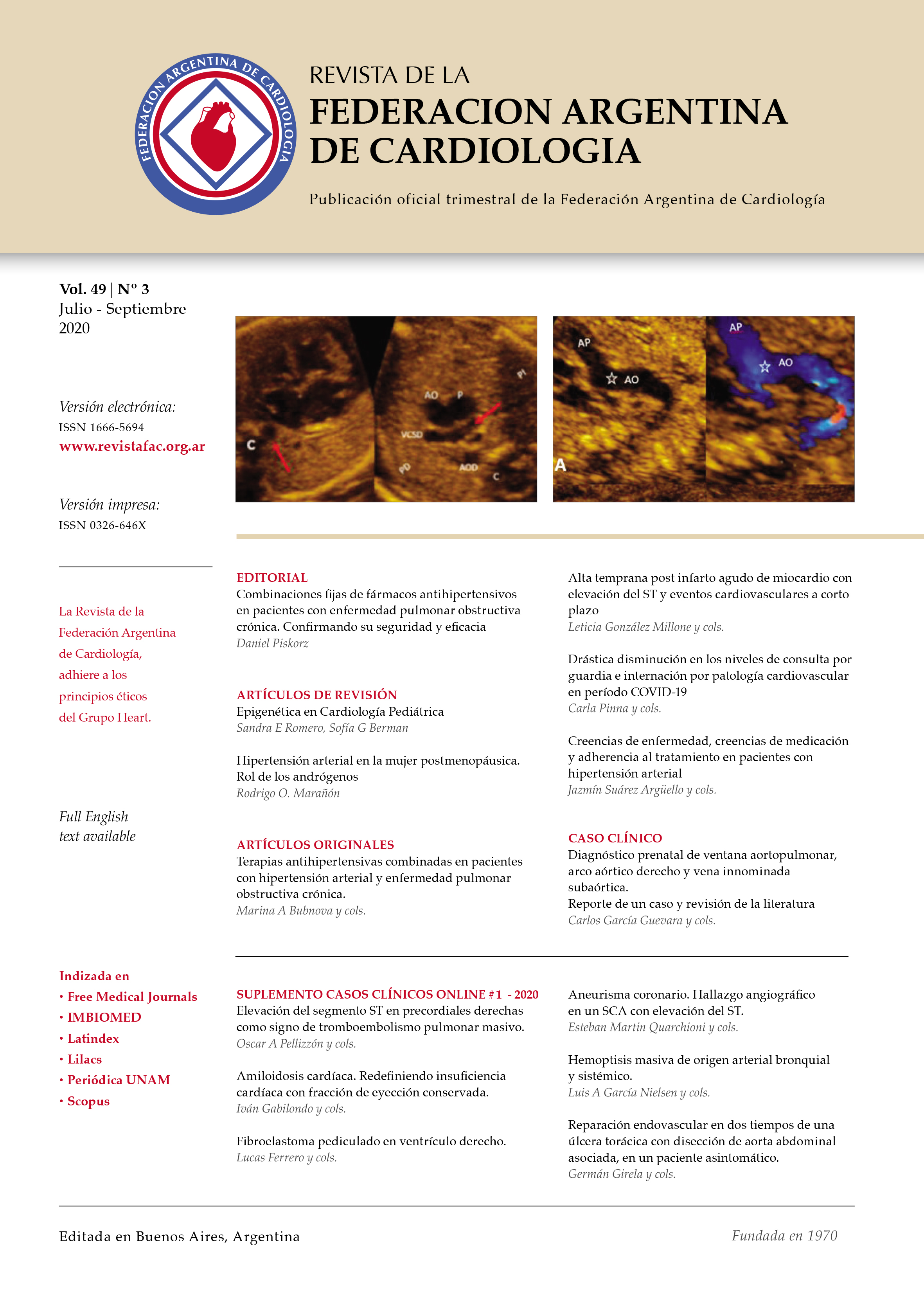
referred to the National Reference Center for Congenital Heart Diseases William Soler Pediatric Cardiocenter. An echocardiographic test was performed, showing the presence of an aortopulmonary window with a right aortic arch and a subaortic innominate vein. The study was performed with a Japanese-made Alpha 10 echocardiogram. Prenatal diagnosis was confirmed at birth, and
an esophageal atresia with distal fistula and one knee dislocation was found. She was operated one day after the birth of the digestive pathology, dying one month later.